Why Your Stomach Doesn't Digest Itself
People are walking contradictions — but every contradiction was tuned. Nature spent billions of years dialing them in. That is the sentence this whole tour starts with.
Take the stomach.
The stomach's acid is hydrochloric acid. At rest, it sits at a pH between 1.5 and 2 — acidic enough to bubble on concrete, enough to dissolve beef, cartilage, and most bacteria on contact.
And the stomach itself? It is a piece of protein. Exactly the kind of tissue that acid eats.
It swims in acid that could digest it, every single day, and it survives. The trick is a layer of jelly.
The Acid Barrier Thinner Than a Millimeter
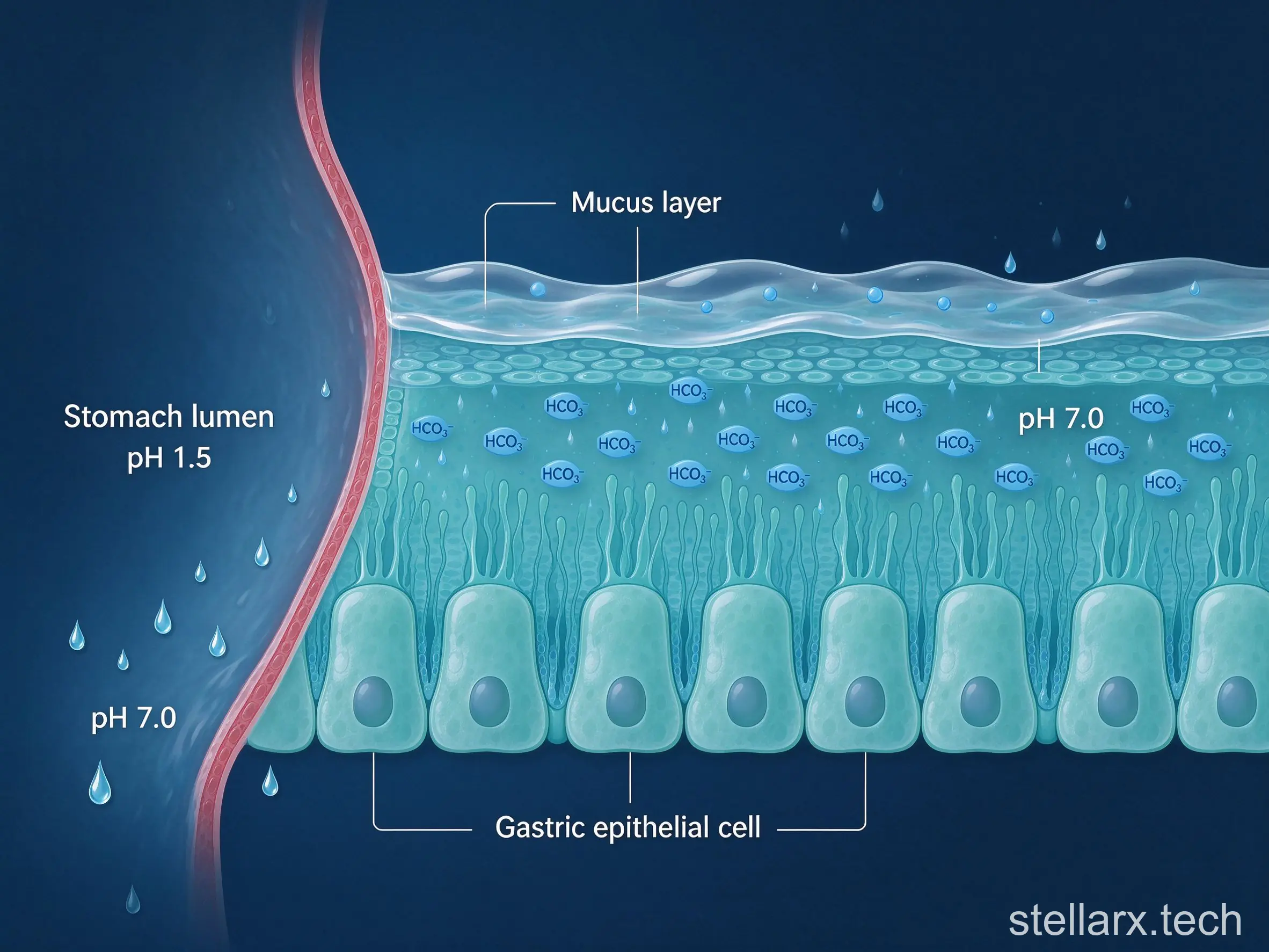
The gastric wall is lined with mucus — a gel layer only fractions of a millimeter thick. Beneath it, the wall's cells constantly pump bicarbonate ions against the concentration gradient, up toward the mucus surface. Bicarbonate: the same thing as baking soda.
Inside that thin jelly, a steep microscopic gradient forms. The side facing the stomach cavity stays strongly acidic at about pH 1.5. The side pressed against the cells is neutralized to near pH 7.0.
A living person maintains an acid-versus-neutral safety line under a millimeter thick, inside their own abdomen, every single day.
And the lining itself is aggressively replaced. The gastric surface epithelium turns over completely every two to three days. The stomach lining you grew last week has already been flushed out of your own digestive tract as metabolic waste.
Different foods take very different times to leave the stomach — liquids pass in tens of minutes, while heavy meals can stay for hours.
| Food | Time in the stomach (minutes) |
|---|---|
| Liquids | 10–60 |
| Fruit | 20–40 |
| Vegetables | 30–60 |
| Dairy | 90–300 |
| Nuts | 120–180 |
| Fish | 30–60 |
| Chicken | 90–120 |
| Beef / lamb | 180 |
| Pork | 270 |
| Pasta | 240 |
| Sandwich | 270 |
| Bento box (mixed meal) | 350 |
Why the World Never Goes Dark, Even When You Stare
Second design: the eyes.
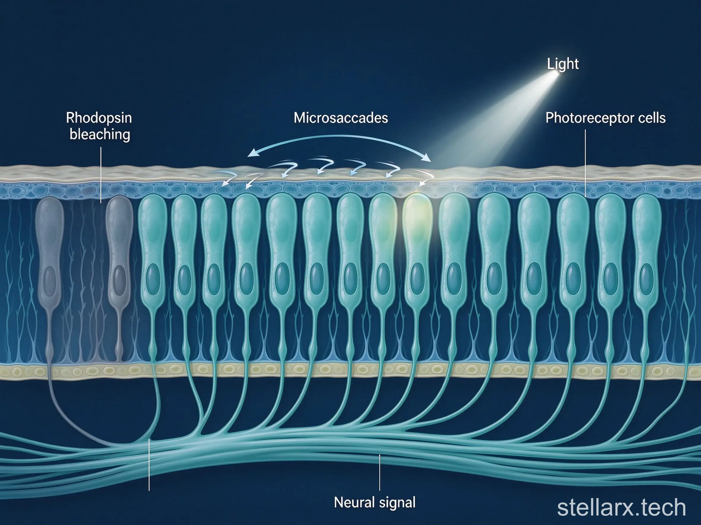
Point a camera at a white wall and the sensor keeps outputting frames. Nerve cells do not work like that. If photoreceptors on the retina receive one constant intensity of light, rhodopsin bleaches rapidly and the neural signal fatigues within seconds.
If your eyeball were perfectly still, staring at a fixed newspaper, the whole visual field would fade within five seconds — and then go pure black.
Completely still.
But you stare at screens for hours and the world never dims. Because your brain runs six extraocular muscles behind the scenes, making dozens of imperceptible micro-movements every second. These are microsaccades.
Your eyeball trembles at micron-scale amplitude, at high frequency, constantly shifting the projected image onto neighboring photoreceptors. The batch of cells that just fatigued is relieved by the next batch, relay after relay, until the visual cortex stitches together a stable, continuous, never-fading picture.
You think you see a still world. In fact, your eyeballs are twitching dozens of times per second at microscopic scale, outrunning the nervous system's own dulling mechanism.
Like a gimbal — holding your eyes at a constant refresh rate.
Six muscles do the work, and each one moves the eye in a different direction.
| Extraocular muscle | Main action |
|---|---|
| Medial rectus | Adducts — turns the eye inward |
| Lateral rectus | Abducts — turns the eye outward |
| Superior rectus | Elevates — rolls the eye upward |
| Inferior rectus | Depresses — rolls the eye downward |
| Superior oblique | Depresses and intorts the eye |
| Inferior oblique | Elevates and extorts the eye |
| Levator palpebrae superioris | Lifts the upper eyelid |
| Six extraocular muscles steer the eyeball; microsaccades are their constant micro-adjustments, dozens per second. | |
Bones Are a 3D Printer Running in Reverse
Third: bones.
Many people believe adult bones are like steel-reinforced concrete — formed once, then a lifeless dead thing. Some even assume fractures do not hurt that much.
But physiologically, bone is a smart load-bearing structure doing reverse 3D printing for life.
The trabecular mesh inside bone is a mix of collagen and hydroxyapatite crystals. When bone bends under mechanical stress, the crystals produce a weak piezoelectric voltage. Nearby bone cells pick it up precisely.
Where stress lands year after year, osteoblasts accelerate matrix secretion, thickening and densifying the trabeculae along the loading direction. Where no load arrives, osteoclasts act like a demolition crew, dissolving and absorbing the excess bone.
CT-scan a professional tennis player and you will see it: the bones of the racket arm are visibly thicker than the other arm, and the internal trabecular alignment follows the line of force from the swing.
The body is constantly dismantling idle bricks and moving them to reinforce the foundations that take impact. It wastes not a single gram of extra weight.
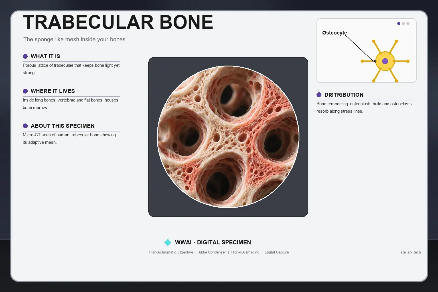
That mesh is not uniform. It is a layered structure that can be seen at different scales, from the millimeter mesh down to nanometer crystals.
| Scale bar | What becomes visible |
|---|---|
| 200 µm | The porous mesh of trabecular bone — the sponge-like lattice |
| 50 µm | Interwoven fibers and the detail of the pores |
| 200 nm | Granular, plate-like mineral particles |
| 20 nm | Rod- and needle-shaped hydroxyapatite crystals |
A Network That Stays Liquid Until It Must Become a Plug
Fourth: the blood-clotting system.
More than ten thousand kilometers of blood vessels hold a few liters of blood, which must stay perfectly fluid. One sticky blockage anywhere means a heart attack or a stroke.
But cut your finger on a piece of paper, and within seconds the damaged site triggers a coagulation cascade that amplifies thousands of times — commanding platelets and fibrin to weave a hard mesh over the wound.
The most precise part: this aggressive clotting response is locked into a few microns around the damage by anticoagulant molecules secreted by the vascular endothelium. The leak is plugged. The main highway keeps flowing.
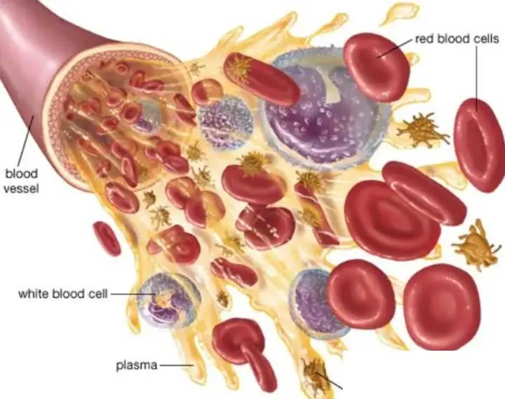
The blood itself carries the pieces of this system — cells and plasma that each do their own job.
| Blood component | Role |
|---|---|
| Red blood cells | Carry oxygen through the body |
| White blood cells | Defend against infection |
| Platelets | Form the clot plug with fibrin when a vessel is damaged |
| Plasma | The liquid matrix that carries cells and clotting factors |
A Decentralized Loop, Tuned Over Billions of Years
No organ, enzyme, or neural circuit in this body takes orders from the top. Each one emerged from billions of years of micro-adjustment and trial — a self-consistent, decentralized closed loop.
You are tuned. So do not change your body's parameters casually.
Looking Closer at the Machinery
Every structure in this tour — the stomach wall, the eye, the bone, the blood — is tissue you could hold on a glass slide. The bone story ends where the microscope begins: trabecular bone, collagen and crystals woven together, is a specimen you can actually look at.
Ever wanted to see cartilage or bone tissue with your own eyes — not in a diagram, but under real optics? WWAI puts a digital specimen library in your pocket, including a Human Hyaline Cartilage specimen where the glassy matrix and rounded chondrocytes are clearly visible. Zoom from 4X to 100X, and ask the AI anything about what you are seeing. Search WWAI in your app store and take the tour yourself.

Related reading

Anticipatory Homeostasis: How Your Body Foresees
Why Stomach Acid Can't Kill Parasite Eggs
Runner's High: Why a Brutal Leg Day Rocks
References
Martinez-Conde S, Macknik SL, Hubel DH. The role of fixational eye movements in visual perception. Nat Rev Neurosci. 2004;5(3):229-240.
Wolff J. Das Gesetz der Transformation der Knochen. Berlin: A. Hirschwald; 1892.
Davie EW, Ratnoff OD. Waterfall sequence for intrinsic blood clotting. Science. 1964;145(3638):1310-1312.
Flemström G, Garner A. Gastroduodenal HCO3− transport: characteristics and proposed role in acidity regulation and mucosal protection. Am J Physiol. 1982;242(3):G183-G193.
- Can a Bunny Conquer the Jianghu? AI Animation Sparks Zootopia Debate
- 當「兔小卯」遇上江湖夢:AIGC動畫《劍兔江湖》掀起萌寵武俠熱潮
- Love Across Distance: Netizens Rally Against Heartbreak in AI Short Drama
- 熱戀隔天就分離?AI短劇《別躲》引發「防刀」焦慮與純愛呼喚
- AI Reimagining of ‘Pet Shop of Horrors’ Sparks Debate: Fans Demand Restoration of Count D’s Iconic Androgyny
- AI重現《恐怖寵物店》引熱議:D伯爵形象崩壞?極樂鳥悲劇喚醒人性反思
- AI-Generated K-Drama Short Series Sparks Debate Over Identity and Aesthetics
- AI短劇爆紅?「韓系穿越」語言混亂與臉孔崩壞引發全網熱議
- Ancient Drama’s Fertility Twist Sparks Debate on Marriage and Modern Storytelling
- 古裝短劇「前夫不孕我另嫁」爆紅:是女性覺醒爽文,還是生育焦慮的市場試探?

全部评论