New Microscope Slides: Four Ways to See Life
A single cell pinches into two. A leaf surface hides tiny openings. Another leaf contains chambers much larger than the cells around them. The smallest structures can give you the clearest place to start looking.
New specimens have arrived in Wwai. Here are four highlights from the latest additions, chosen for a simple reason: each gives you a distinctive structure to find, rather than another name to memorise.
Paramecium binary fission: find the narrowing waist
A familiar slipper-shaped outline takes an unfamiliar turn in the Paramecium binary fission specimen. The body is constricted across its middle, preserving a stage in which one cell is becoming two. Start with that narrowing waist, then follow the outlines of the two developing halves.
This is transverse division: the constriction crosses the short axis of the elongated cell. The specimen turns “a single-celled organism reproduces by division” into a shape you can inspect. Your first observation challenge is to locate the connection between the halves before looking for finer internal details.
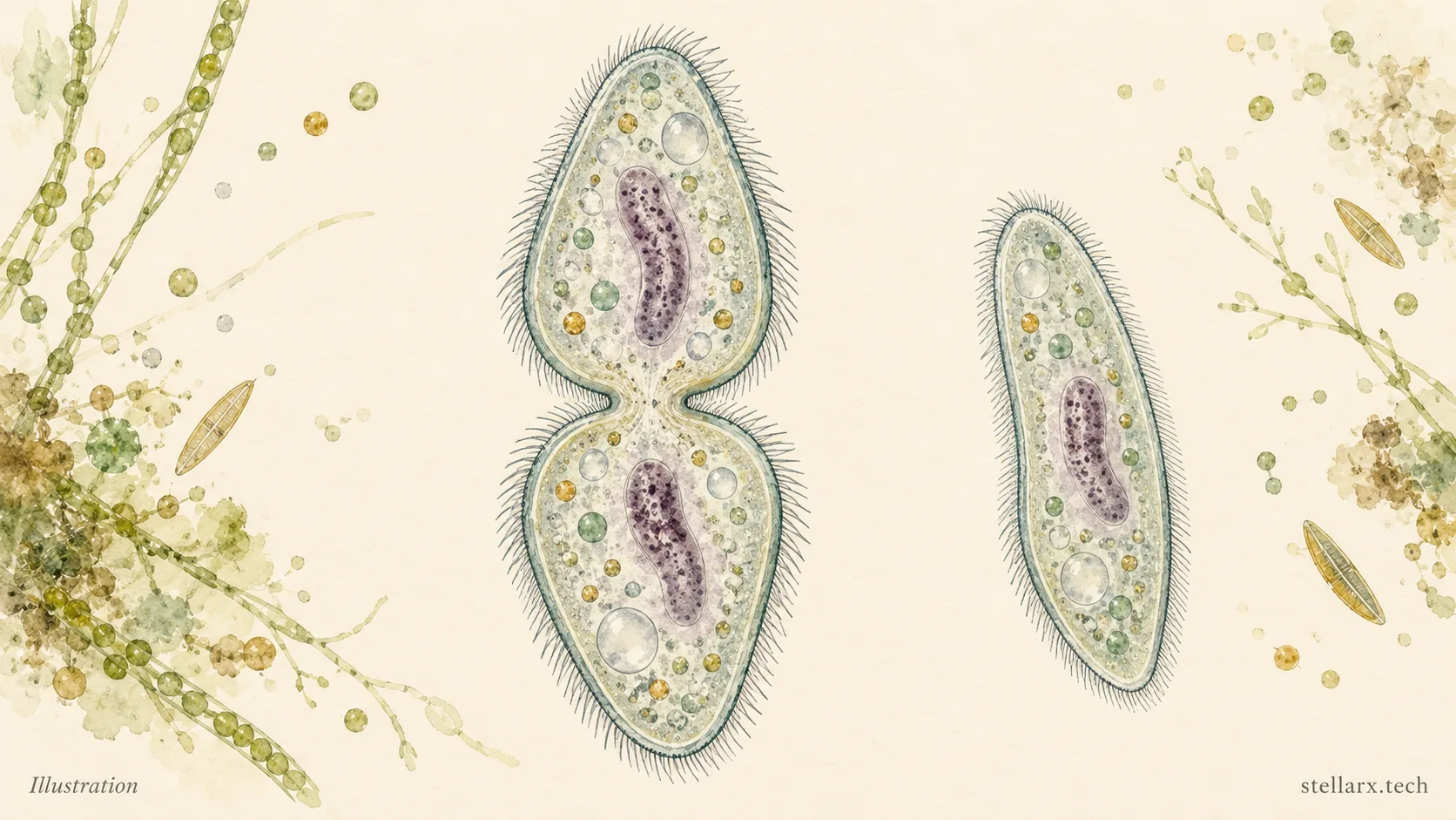
Illustration: a dividing Paramecium and an undivided form for comparison.
Spinach leaf epidermis: find a pair among the puzzle pieces
The spinach leaf epidermis specimen introduces a different kind of pattern. Its surface cells have wavy, interlocking boundaries. Among these puzzle-like outlines, look for two curved guard cells surrounding a small opening: a stoma.
Guard cells regulate the pore through which gases and water vapour pass. In this specimen, chloroplasts within the guard cells provide another detail to look for. First trace one complete pair; then find another nearby and compare it with the larger, irregular surrounding cells.
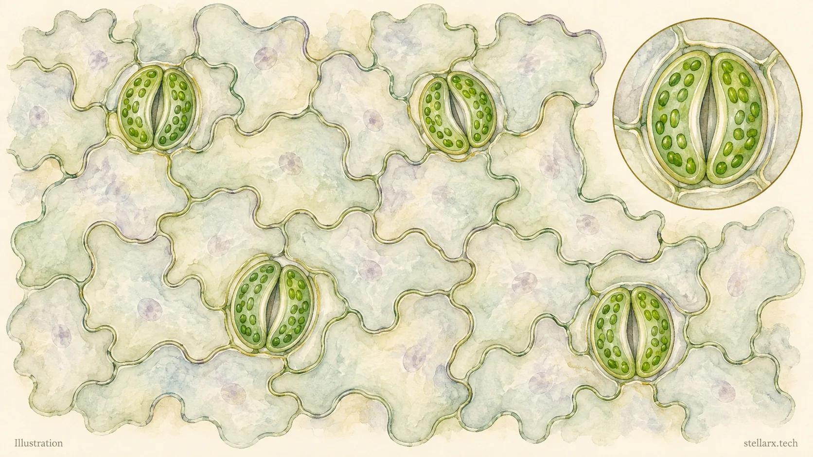
Illustration: a surface view of leaf epidermal cells and stomatal complexes.
Corn root tip: follow the changing cell shapes
The corn root tip longitudinal section lets you look along a growing root. Begin at the protected tip and follow the cell files upward. Small, closely packed cells give way to longer cells farther from the tip; the specimen description also identifies the root cap, meristematic region, elongation region and mature region.
Instead of trying to label every cell at once, compare two areas. Where are cells shortest and most tightly packed? Where do they become elongated? The contrast gives the familiar word “growth” a visible organisation: different regions contribute in different ways.
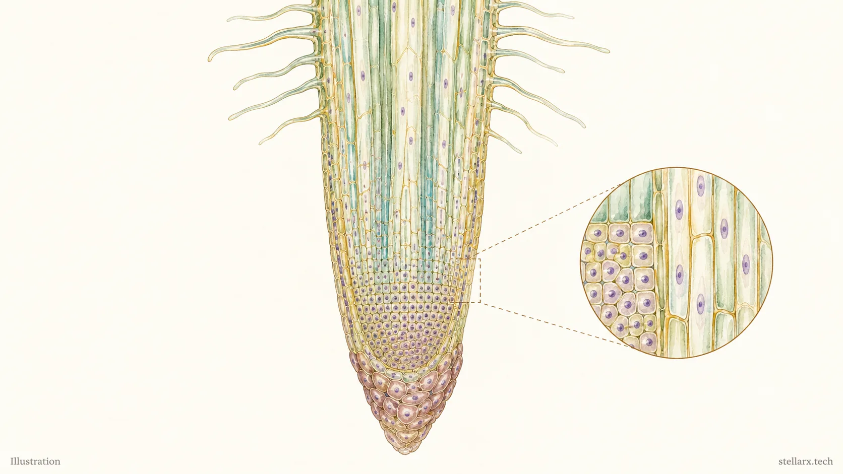
Illustration: a simplified root tip with small cells near the tip and elongated cells farther above.
Water lily leaf: look at the spaces between cells
In the water lily leaf cross section, the most striking feature is the open space. Large internal air chambers interrupt the tissue, with cells forming partitions around them. These conspicuous spaces are part of aerenchyma, the air-space-rich tissue characteristic of many aquatic plants.
Follow the upper and lower boundaries of the leaf, then look inward. Compare the size of an air chamber with the cells surrounding it. After the tightly packed root tip, this leaf offers a useful change of perspective: an empty-looking region can be part of the structure worth observing.
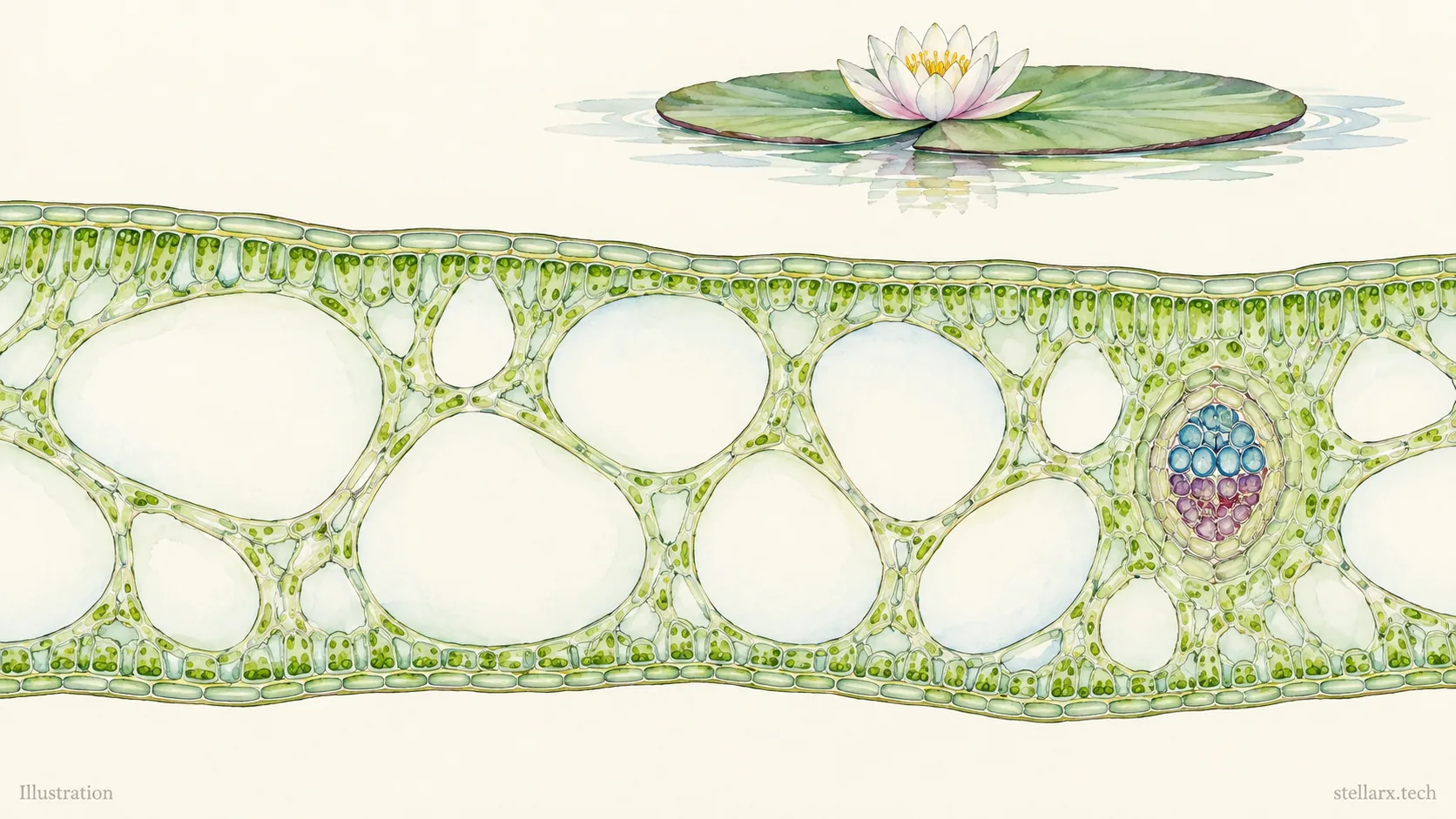
Illustration: a simplified floating leaf cross section highlighting internal air spaces.
Choose one specimen and one question
Start with the specimen that catches your eye. Find the constriction, the guard-cell pair, the change in cell length or the air chamber. Then move to another specimen and ask what changed. Four preparations offer four different ways to practise observing, from a whole single-celled organism to the architecture inside a leaf.
Ready to explore the new arrivals? Search “Wwai” in your app store and download or update to Wwai 1.0.4 to experience the new specimens. The Paramecium binary fission specimen is a good first stop: begin at the waist, then let the details lead you further.

References
[1] OpenStax. Biology 2e, “Groups of Protists.” https://openstax.org/books/biology-2e/pages/23-3-groups-of-protists
[2] OpenStax. Biology 2e, “Leaves.” https://openstax.org/books/biology-2e/pages/30-4-leaves
[3] OpenStax. Biology 2e, “Early Plant Life.” https://openstax.org/books/biology-2e/pages/25-1-early-plant-life
[4] University of Florida, IFAS. “Cell Types: Parenchyma.” https://propg.ifas.ufl.edu/01-biology/02-cell-types/07-celltypes-parenchyma.html
- 凉土豆能减肥?先看看土豆的细胞
- New Microscope Slides: Four Ways to See Life
- 顯微鏡標本上新:四個看見生命細節的起點
- The Pig Paradox: High Intelligence, Low Self-Esteem
- 豬的聰明與悲歌:當高智商遇上餐桌,我們該如何自處?
- The Silent Pain: Why Your Cat’s “Picky Eating” Might Be a Dental Emergency
- 貓咪「挑食」竟是牙根被吃空?揭開高隱蔽性牙吸收病,別讓毛孩默默忍痛
- The Paradox of Pet Birds: Legal Ambiguity and the Rise of the Monk Parakeet
- A Barber Communication Card Is the Next Planned Feature
- 鸚鵡飼養灰色地帶:科普熱度下的法規爭議與萌寵焦慮
全部评论