How Kidneys Work: Filter, Reabsorb, Keep What Matters
Your two kidneys receive about 1,200 mL of whole blood every minute — 20 to 25 percent of your cardiac output. Each day, the glomeruli filter roughly 180 liters of primary urine, and only about 1.5 liters leave the body as final urine.
That means more than 99 percent of the water, and nearly every nutrient, is precisely reabsorbed back into the blood.
Calling the kidney a "sieve" only explains how it filters blood; it cannot explain how the organ manages to expel waste while holding on to almost everything valuable. A better image: the kidney is a highly automated recycling plant. It first does a coarse filtration, then uses a series of molecular pumps and channels along a long pipeline to collect every useful thing back into the blood, finally letting only true waste and a little excess water go.
The Nephron: The Kidney's Work Unit
The core workplace of the kidney is the nephron. Each kidney contains about one million of them.
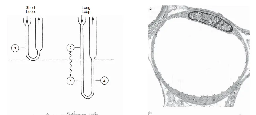
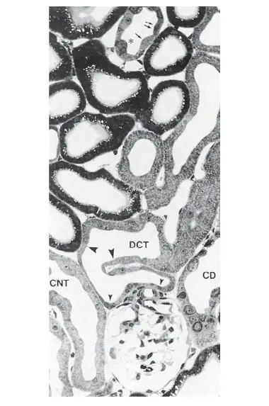
Every nephron has two parts: the renal corpuscle (the glomerulus plus Bowman's capsule) and the tubular system — the proximal tubule, the loop of Henle, the distal tubule, and the collecting duct.
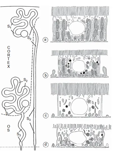
Waste excretion and water balance run through three core stages: glomerular filtration (coarse selection, forming primary urine), tubular reabsorption (fine selection, recovering nutrients and water), and tubular secretion and excretion (supplementary clearance and acid-base regulation).
The Glomerular Filter: A Triple-Check "Molecular Customs"
Blood enters the kidney through the afferent arteriole, which branches into the capillary network of the glomerulus. Water and small solutes are pressed into Bowman's capsule to form primary urine. The glomerulus can "let metabolic waste and small molecules pass while forbidding red blood cells and large proteins" thanks to a three-layer filtration membrane.
Inner layer — capillary endothelium. The endothelial cells carry fenestrations about 70–90 nm across. This acts as a coarse screen: it blocks blood cells (red cells are about 7.5 µm) but lets plasma proteins and all small molecules pass freely.
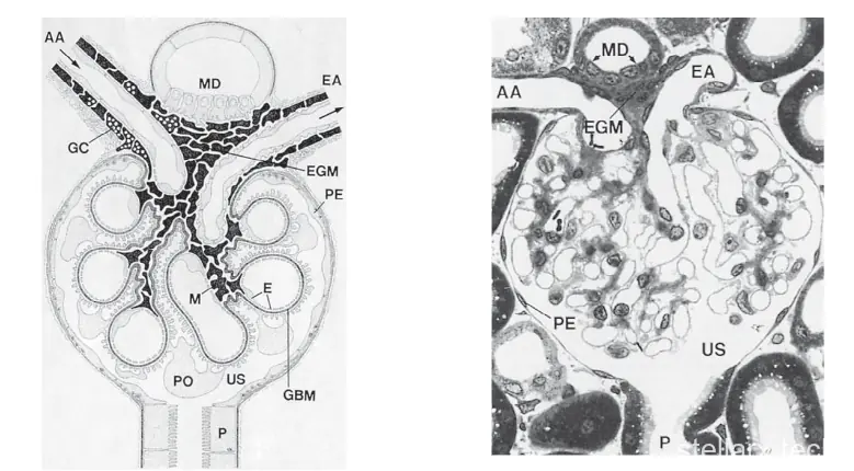
Middle layer — glomerular basement membrane. Built from type IV collagen, laminin, and heparan sulfate proteoglycans, it is about 300 nm thick. It acts as a mesh barrier with pores of about 2–8 nm, effectively stopping most mid- and large-sized proteins such as albumin (≈68 kDa, radius ≈3.6 nm).
Outer layer — podocytes and their slit membranes. Podocytes attach to the outer face of the basement membrane and extend interlocking primary and secondary foot processes, separated by narrow slits bridged by dedicated slit membranes. The main proteins are nephrin and podocin. The effective slit width is only about 4–11 nm — the final, most critical barrier against protein leakage.
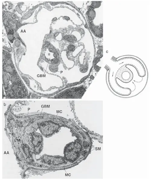
Size and Charge: A Double Barrier
Filtration selectivity does not rely on molecular size alone (the mechanical barrier); there is also a charge barrier. The basement membrane and podocyte surfaces are rich in negatively charged glycans such as heparan sulfate. The main plasma proteins, including albumin, also carry a net negative charge at physiological pH. Like charges repel: negatively charged albumin finds it extremely hard to cross the filter.
Judged by size alone, albumin (≈69,000 Da, radius ≈3.6 nm) should be able to squeeze through in part. Yet normal urine contains almost no albumin. The reason: albumin carries a large net negative charge at physiological pH, while every layer of the glomerular barrier — the endothelial surface, the basement membrane, and the podocyte foot processes — is coated with negatively charged glycoproteins. These fixed negative charges form an electrostatic repulsion field. Negatively charged albumin gets pushed back as it approaches, while positively charged molecules pass more easily. The glomerulus's logic: small molecules and neutral/positive small molecules pass freely; large molecules and negatively charged large molecules are blocked. This dual selectivity ensures waste is filtered out while plasma proteins — things that must not be lost — are retained as completely as possible.
Dynamics: Effective Filtration Pressure
Primary urine production is a contest of physical pressures.
| Effective filtration pressure (EFP) | Value / role |
|---|---|
| EFP = Pcapillary − (Pcapsule + πcapillary) | Net pressure driving filtration |
| Glomerular capillary pressure (Pcapillary) | ≈ 45–50 mmHg — the driving force (efferent arteriole resistance keeps the glomerulus under high pressure) |
| Bowman's capsule pressure (Pcapsule) | ≈ 10–15 mmHg — opposes filtration |
| Plasma colloid osmotic pressure (πcapillary) | ≈ 20–35 mmHg (rising along the capillary) — pulls water back into vessels, opposes filtration |
| Filtration equilibrium | When EFP = 0, filtration stops (proteins concentrate as water leaves) |
Tubular Reabsorption and Secretion
Primary urine contains no blood cells and very little protein; otherwise its concentrations of water, glucose, amino acids, electrolytes, and metabolic waste (urea, creatinine) match plasma exactly. Without reabsorption, the body would dehydrate and collapse within hours. The tubule recovers everything useful through precisely engineered transporter proteins.
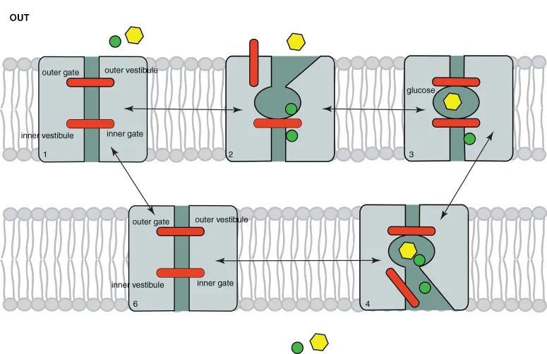
1. Proximal tubule — the main recovery worker. The proximal tubule does the heaviest work: about 65–70 percent of Na⁺, Cl⁻, and water, 80 percent of HCO₃⁻, and almost all glucose and amino acids are reabsorbed here. The core engine is the Na⁺/K⁺-ATPase on the basolateral membrane. It continuously pumps Na⁺ out of the cell into the interstitial fluid, keeping intracellular Na⁺ low and creating a Na⁺ gradient at the apical membrane. Sodium flows down this gradient into the cell, carrying glucose and amino acids with it through SGLT2/SGLT1 (sodium-glucose cotransporters) and Na⁺-amino acid cotransporters. Glucose and amino acids then leave through the basolateral membrane and return to the blood.
Water reabsorption is passive: as Na⁺ and solutes enter the interstitial fluid, local osmolality rises, and water follows the osmotic gradient through the paracellular pathway and the transcellular pathway (AQP1 water channels) into the interstitial fluid, then into peritubular capillaries. Solutes are recovered and water comes along.
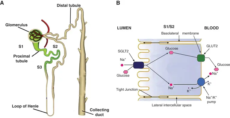
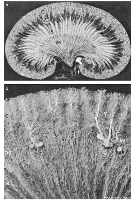
2. Loop of Henle — the countercurrent multiplier. The loop's special job is not to reabsorb large amounts of water directly but to build a hyperosmotic environment in the renal medulla, preparing the collecting duct to concentrate urine. The thin descending limb is permeable to water (via AQP1) and nearly impermeable to solutes. The thick ascending limb is impermeable to water but actively pumps Na⁺, K⁺, and Cl⁻ into the medullary interstitium via NKCC2 (sodium-potassium-2 chloride cotransporter). Because the descending and ascending limbs carry fluid in opposite directions (countercurrent), this pumped NaCl keeps "multiplying" the deep-medullary osmolality, building a gradient from cortex to medulla — from about 300 mOsm/kg in the cortex to 1,200 mOsm/kg deep in the medulla. The thick ascending limb is water-impermeable, so tubular fluid is "diluted" while the medullary interstitium is "concentrated." The drug furosemide (Lasix) targets exactly this NKCC2.
3. Distal tubule and collecting duct — the fine-tuning final valve. By the distal convoluted tubule and collecting duct, about 90 percent of the filtrate has been reclaimed; the rest is regulated with great precision under hormonal control. The distal tubule reabsorbs about 5–8 percent of Na⁺ and Cl⁻ via NCCT (sodium-chloride cotransporter), remaining water-impermeable and diluting the tubular fluid further. Thiazide diuretics target this NCCT. Collecting duct principal cells reabsorb Na⁺ through ENaC (epithelial sodium channel) and secrete K⁺. Critically, the collecting duct's water permeability is controlled by antidiuretic hormone (ADH).
4. Zero-loss protection for nutrients — secondary active transport. Take glucose: it is reabsorbed 100 percent in the proximal tubule. The energy source is the Na⁺/K⁺-ATPase on the basolateral membrane, which keeps intracellular Na⁺ extremely low and maintains a negative membrane potential. At the apical membrane, SGLT2 uses the huge Na⁺ electrochemical gradient to pull one Na⁺ and one glucose molecule into the cell together (secondary active transport). On the basolateral side, high intracellular glucose diffuses down its gradient through GLUT2 carriers into the interstitial fluid and onward into capillaries. The renal threshold: proximal tubule glucose reabsorption has an upper limit. When blood glucose exceeds about 8.9–10.0 mmol/L (160–180 mg/dL), filtered glucose outruns the SGLT transport capacity, and the excess spills into the urine — the glucosuria of diabetes, which also drives osmotic diuresis.
5. Water recovery and concentration — the countercurrent system. The kidney must also match hydration state, producing concentrated or dilute urine. This depends on the medullary countercurrent multiplier: NKCC2 on the thick ascending limb pumps Na⁺ and Cl⁻ into the medullary interstitium while that segment stays water-impermeable, leaving deep medullary osmolality extremely high. When primary urine flows through the water-permeable descending limb and collecting duct, the hyperosmotic medulla acts like a sponge, pulling water back into the body via AQP1 and AQP2. The U-shaped vasa recta run with very slow flow — absorbing solutes and releasing water on the way down, absorbing water and releasing solutes on the way up — so they carry away reabsorbed water and solutes without destroying the medullary gradient.
6. Precise fluid balance — aldosterone and ADH. When the body dehydrates and plasma crystalloid osmolality rises, the hypothalamus produces ADH (vasopressin), released by the posterior pituitary. ADH binds V₂ receptors on the basolateral membrane of collecting duct principal cells, activates the cAMP pathway, and triggers vesicles containing aquaporin-2 (AQP2) to fuse into the apical membrane — greatly increasing water permeability and concentrating the urine. When blood volume is low or potassium is high, the renin-angiotensin-aldosterone system (RAAS) activates: aldosterone acts on the distal tubule and collecting duct to induce more ENaC and Na⁺/K⁺-ATPase, achieving "save sodium, save water, excrete potassium."
When the Kidneys Fail: Peritoneal Dialysis
When severe disease (chronic glomerulonephritis, diabetic nephropathy) destroys nephrons, end-stage renal disease (ESRD, uremia) follows: metabolic waste (urea, creatinine) and excess water cannot be excreted, and acid-base and electrolyte balance breaks down. Replacement therapy is needed — and peritoneal dialysis ingeniously uses the body's own biological membrane to mimic kidney function.
1. The peritoneum as a natural dialysis membrane. The abdominal cavity is lined by a semipermeable membrane, the peritoneum, with a surface area comparable to body surface area (about 1.5–2.0 m²), richly supplied with capillaries. Its anatomy resembles the glomerular filtration membrane, with three layers: the vascular endothelium and basement membrane, the peritoneal interstitium (a gel of collagen network and glycosaminoglycans), and the mesothelial cell layer (a single-layer flattened epithelium with microvilli).
2. Core physics of peritoneal dialysis. Clinically, sterile dialysis fluid is instilled into the abdominal cavity and left for several hours. Plasma and dialysate exchange across the peritoneum:
① Solute removal — diffusion. The dialysate contains no urea or creatinine. Driven by concentration gradients, blood urea nitrogen, creatinine, and excess potassium diffuse across the peritoneum into the dialysate.
② Water removal — ultrafiltration (osmosis). Peritoneal dialysis has no high mechanical blood pressure to create an "effective filtration pressure." To pull excess water out, the dialysate carries a high concentration of osmotic agent — most commonly glucose (1.5%, 2.5%, or 4.25% glucose dialysate). High glucose raises dialysate osmolality (346–485 mOsm/kg) well above plasma (≈285 mOsm/kg), and the osmotic difference draws water from blood into the abdominal cavity.
③ Acid-base correction — buffer supply. The dialysate contains lactate or bicarbonate at high concentration; buffer diffuses down its gradient into the blood, correcting the metabolic acidosis common in uremia.
3. Kidney physiology vs peritoneal dialysis, side by side:
| Mechanism | Human kidney (natural) | Peritoneal dialysis (artificial) |
|---|---|---|
| Driving force | Blood pressure from the heartbeat (hydrostatic pressure gradient) | High osmotic gradient from glucose or polyglucose |
| Semipermeable membrane | Endothelial cells + basement membrane + podocyte slit membrane | Peritoneal capillary endothelium + interstitium + mesothelial layer |
| Solute clearance | Convection (filtration): solutes carried out with bulk fluid flow | Diffusion: solutes move down their chemical concentration gradients |
| Water handling | Reabsorbed via medullary osmotic gradient and AQP1/AQP2 channels | Pulled out by dialysate osmolality and AQP1 |
| Protein retention | Mechanical + charge barrier; protein filtration fraction < 0.01% | Size barrier only; still loses ~5–10 g albumin per day |
The Elegance of the Kidney
The kidney is not a simple filter at all. It is a dynamic balancing system that first filters everything and then precisely reabsorbs and secretes: coarse filtration at the glomerulus (three-layer barrier plus hydrostatic pressure, pushing everything except cells and large proteins into the tubule — the charge barrier is what keeps albumin from leaking, something no sieve metaphor can explain); fine recovery in the tubule (using the Na⁺/K⁺-ATPase electrochemical gradient and secondary active transport to reclaim 100 percent of glucose, and the medullary countercurrent system plus water channels to reclaim 99 percent of water); regulation by hormones (ADH and aldosterone adjusting dynamically to maintain internal stability); and secretion and excretion (the tubule actively secretes H⁺, K⁺, and NH₃, participating in acid-base and electrolyte balance).
When kidney function fails, peritoneal dialysis reproduces, at the macroscopic level, the kidney's filtration and waste-removal physics through high-osmotic glucose gradients and solute diffusion — a meeting point of biology and clinical medicine.
A sieve can only filter; it cannot collect. The greatness of the kidney is that it filters while keeping almost everything that should be kept.
The same principle hides in the microscopic world: a tissue is a system of precisely organized tubes and cells, each doing its own job — visible only under a microscope. With WWAI, you can see them the moment curiosity strikes, including a female ascaris cross-section specimen where the layered body-wall and internal tubular organs are clearly visible. Search "WWAI" in your app store and download it today.

References
Haraldsson, B. & Jeansson, M. (2008). Properties of the glomerular barrier and mechanisms of proteinuria. Physiological Reviews, 88(2), 451–487.
Kanai, Y. et al. (1994). The human kidney low affinity Na+/glucose cotransporter SGLT2. Journal of Clinical Investigation, 93(1), 397–404.
Weitzman, R.E. & Kleeman, C.R. (1979). The clinical physiology of water metabolism. Part II: Renal mechanisms for urinary concentration; diabetes insipidus. Western Journal of Medicine, 131(6), 486–515.
Vallon, V. & Thomson, S.C. (2020). The tubular hypothesis of nephron filtration and diabetic kidney disease. Nature Reviews Nephrology, 16(6), 317–336.
Hasler, U. et al. (2009). Aquaporin-2 abundance in the renal collecting duct. American Journal of Physiology - Renal Physiology, 297(1), F10–F18.
Lindholm, B. et al. (1987). Kinetics of peritoneal dialysis with glucose-based fluids. Peritoneal Dialysis Bulletin (kinetics study).
Figure credits: physiology and histology figures reproduced from the original source article for educational science communication; locally adapted for this site's science-communication positioning.
Related reading

Kidney Function: How Waste Leaves the Body


- 同手性是什麼?2026 諾貝爾化學獎得主解開生命之謎
- Homochirality Explained: Nobel Chemistry 2026 | stellarx
- Is Using Your Phone in Bed Bad for Your Eyes?
- AI Consumer Spending: Who Actually Pays for AI?
- How Do Bees Communicate? Dance and Pheromones
- How Kidneys Work: Filter, Reabsorb, Keep What Matters
- Female Thigh Fat: A Second Sex Characteristic
- IceCube Neutrino Detector: Ghost Particle Hunter | stellarx
- Lobster Mushroom: The Fungus That Parasitizes Mushrooms
- Sacculina: The Crab-Castrating Parasite Explained
全部评论