How the Kidney Filters 180 Liters of Plasma a Day
In 1943, a Dutch doctor built a machine out of sausage casing, an orange-juice can, and scrap car parts. He used it to treat 16 kidney-failure patients. The first 15 died. The 16th — a 67-year-old woman — became the first person in history saved by an artificial kidney.
The organ he was trying to replace does something stranger than any machine: every day it filters 180 liters of plasma out of your blood, recycles 99% of it, and sends less than 2 liters of waste down the drain. This is how that impossible pipe system works.
Why the Kidney Can't Just Throw Everything Out
Your body produces metabolic waste around the clock: protein breaks down into urea, muscle activity makes creatinine, nucleic acids make uric acid, plus surplus potassium ions, hydrogen ions, and drug metabolites.
These wastes are too different in size and chemistry for any single transporter protein to recognize them all. So the kidney does something counterintuitive: it filters out every small molecule first, then uses dozens of transporter proteins to haul the useful ones back, one by one. Whatever gets reclaimed is what the body needs. The rest becomes urine.
The Filter: Three Layers, Zero Blood Cells
The basic working unit is the nephron — about a million per kidney. Each one is a complete pipeline from filtering to reclaiming to excreting.
The first step happens in the glomerulus: a ball of capillaries wrapped in a cup-shaped structure (Bowman's capsule). Blood pressure squeezes water and small molecules out of the capillaries and into the cup. Whether a molecule gets through depends on a three-layer filter:
The capillary endothelium has pores of 70–100 nanometers — blood cells can't pass, dissolved small molecules can.
The basement membrane is 300–350 nanometers thick, woven from collagen and glycoproteins, and negatively charged — it repels the likewise-negative plasma albumin.
The podocytes extend slender foot processes that interlock around the capillaries, leaving gaps of about 25–60 nanometers.
Stack them up and anything under roughly 70 kilodaltons gets through, while blood cells and most proteins are held back.
The liquid that emerges is called the primary urine. Its composition is nearly identical to plasma — including glucose at the same concentration as blood, along with amino acids, sodium, and potassium. If it were simply excreted, you'd die within a day from low blood sugar and electrolyte chaos.
SGLT2: The Sodium-Powered Glucose Pump
Most of the heavy lifting happens in the proximal tubule: about 65–70% of the water, almost all the glucose and amino acids, and most of the sodium are pulled back into the blood here.
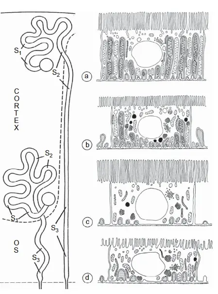
Glucose reclamation depends on a protein called SGLT2. It sits on the lumen-facing surface of the proximal tubule's epithelial cells, almost exclusively in the S1 and S2 segments, where the tubule is broken into segments by cell type [1].
SGLT2 runs on a sodium gradient: sodium concentration is high in the tubule lumen and low inside the cell, so sodium tends to flow inward — and SGLT2 hitches a glucose molecule to each sodium ion it lets in. To keep the gradient alive, a Na⁺/K⁺-ATPase on the blood-facing side pumps sodium out of the cell using ATP. The glucose then exits into the blood through a GLUT2 transporter.
Every day about 180 grams of glucose are filtered. SGLT2 reclaims 90%; SGLT1 in the S3 segment mops up the remaining 10%. That's why normal urine contains no glucose. When blood sugar exceeds the transporters' capacity, glucose spills into urine — the classic diabetes finding. And the modern class of diabetes drugs called SGLT2 inhibitors (like dapagliflozin) work by deliberately crippling SGLT2, so more glucose leaves in the urine and blood sugar falls.
The Loop of Henle: Building a Salt Mountain
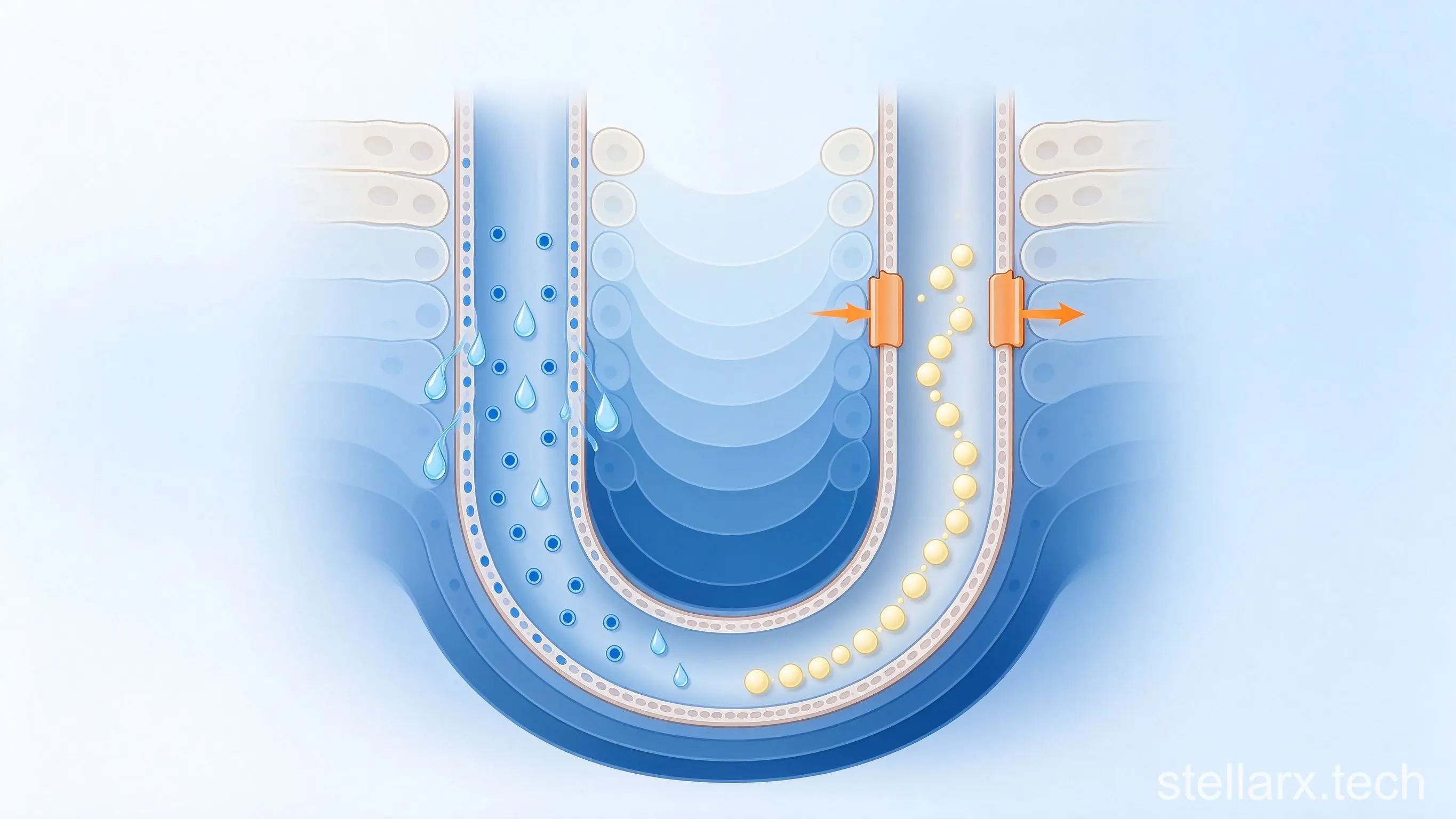
If the kidney stopped here, you'd still urinate 30 to 50 liters of dilute urine every day [2]. It needs a concentration step — and that's the loop of Henle, a U-shaped tube that dips from the cortex into the medulla and folds back.
The two arms of the U behave oppositely. The descending limb is full of water channels: water leaves freely, while sodium and chloride barely cross. The ascending limb is water-tight, but its thick segment pumps Na⁺/K⁺/2Cl⁻ out into the surrounding tissue.
Fluid flows down one arm and up the other while salt is being pumped out sideways — a mechanism called countercurrent multiplication. Each pass raises the salt concentration around the loop, which pulls more water out of the descending limb, which concentrates the fluid further. After several rounds, the deep medulla reaches 1,200 mOsm/L — four times the plasma — the osmotic mountain that makes concentrated urine possible.
Aquaporin-2: The Protein That Decides Urine Color
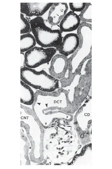
Whether urine actually comes out concentrated is decided by the collecting duct, and specifically by whether its wall carries a water channel called aquaporin-2 (AQP2).
Finding that channel took decades. As early as the late 1800s, researchers noticed that water crossed kidney tubule walls far faster than simple diffusion could explain — something was obviously ferrying it through [3]. But nobody could find the ferryman.
In 1988, at Johns Hopkins, Peter Agre isolated a 28-kilodalton protein while studying red blood cell membranes. He had no idea what it did — he only noticed it was abundant in red blood cells and the kidney. In 1992, he injected the protein's mRNA into Xenopus (African clawed frog) oocytes. Control oocytes floated in hypotonic solution unchanged; oocytes expressing the protein swelled as water poured in and burst within minutes.
He named it aquaporin — Latin for "water pore." A single channel lets billions of water molecules through per second while excluding all solutes — even the smallest, the hydrogen ion — because charged ions are deflected by the electrostatic environment of the channel wall.
In the collecting duct, the position of AQP2 decides everything. When you're dehydrated, the hypothalamus releases antidiuretic hormone (ADH), which makes collecting-duct cells move stored AQP2 into the luminal membrane. Water is reclaimed as fluid passes the deep, salty medulla — urine comes out scanty and dark. When you're well hydrated, ADH drops, AQP2 retreats, the wall becomes water-tight, and urine comes out copious and pale.
That color is worth reading. Here's the standard five-level chart:
| Urine color | What it usually means |
|---|---|
| Clear / colorless | Normal; very well hydrated |
| Pale yellow | Normal; mildly dehydrated |
| Bright yellow | Mildly or moderately dehydrated; or taking dietary supplements (e.g. B vitamins) |
| Amber | Moderately or severely dehydrated |
| Tea / brown | Severely dehydrated — drink water soon |
Back to the Sausage Casing
In the late 1930s, the young doctor Willem Kolff watched kidney-failure patients slowly poison themselves with their own metabolic waste — and could do nothing. He later called it the most helpless period of his life.
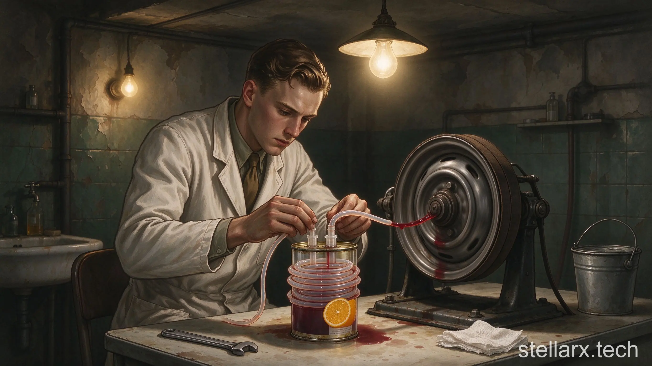
In 1940, he was posted to the small Dutch town of Kampen and started building an artificial kidney. His dialysis membrane was sausage casing made of cellophane, which lets small molecules like urea through but holds back proteins. His container was an orange-juice can. His rotating drum came from scrap car parts.
The first machine was completed in 1943. The first 15 patients died. In 1945, the 16th — a 67-year-old woman — regained kidney function after dialysis: the first verified human survivor of an artificial kidney. After the war, Kolff donated all five of his homemade machines to hospitals around the world. He charged nothing.
Why Dialysis Is Still Only a Slice of the Job
Modern dialyzers have come far from sausage casing, but they still only do one slice of the kidney's job: clearing waste from blood. A real kidney also reclaims glucose and amino acids, balances sodium and potassium, concentrates urine, regulates blood pressure, makes erythropoietin, and activates vitamin D.
A healthy kidney contains at least 51 major cell types [4]. In injured kidney samples, researchers identified 28 distinct cell states — cells mid-repair, cells failing and degenerating, and cells caught in transition. None of that, so far, fits inside a machine.
Ever wondered what that "round, dimpled biscuit" looks like that your glomerulus filters around but never lets through? WWAI is an AI-powered biology encyclopedia with an online microscope — including a red blood cell smear specimen where the doughnut-shaped red cells are clearly visible. Search "WWAI" in your app store and download it today.

Related reading

One Kidney vs Liver Regeneration: Why They Differ

Food Adulteration in Ancient China

Why Thailand's Papaya Salad Is 'Involution' Food
References:
[1] Wright E M. SGLT2 inhibitors: physiology and pharmacology. Kidney360, 2021, 2(12): 2027.
[2] Evans R G. Evolution of the glomerulus in a marine environment and its implications for renal function in terrestrial vertebrates. Am J Physiol Regul Integr Comp Physiol, 2022, 324: R143-R151. doi: 10.1152/ajpregu.00210.2022.
[3] Agre P. Aquaporin water channels (Nobel Lecture). Angewandte Chemie International Edition, 2004, 43(33): 4278-4290.
[4] Lake B B, Menon R, Winfree S, et al. An atlas of healthy and injured cell states and niches in the human kidney. Nature, 2023, 619(7970): 585-594.
All illustrations are AI-generated except where the source image is used; the urine color chart is reproduced as structured HTML data from the source article.
- Your Kidneys Are a Selective Filter You Can Ruin
- Testicular Torsion: The Six-Hour Clock Ticks at Home
- Your Gut Is a Second Brain. It Also Votes on What You Eat
- Prostate Calcification: What the Blip Really Means
- A Daily Bubble Tea Turned This Teen's Neck Black
- Your Gut Makes Most of Your Serotonin
- Raw Spring Water Almost Killed a 61-Year-Old
- Why Restaurant Meals Leave You So Thirsty
- Why Porridge Spikes Blood Sugar Like Soda
- How the Kidney Filters 180 Liters of Plasma a Day
全部评论