Why Some Wounds Heal in Days and Others Never Do
Two cuts of identical size, on the same person, can heal at wildly different speeds. Wound size is only the surface number. What surgeons actually worry about is: can the wound edges come together? Is there tension? Is blood supply adequate? Is there necrotic tissue or foreign material? Are bacteria out of control? Does the body have enough oxygen, protein and immune capacity?
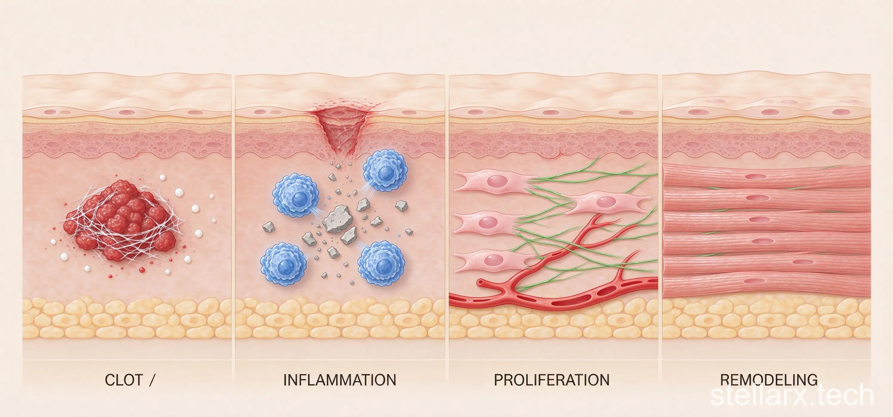
Wound healing is not skin "sticking itself back together." It is a staged repair project: bleeding stops, inflammation cleans up, granulation tissue fills the defect, epithelium covers the surface, collagen remodels. If any stage stalls, the wound drags — or becomes chronic.
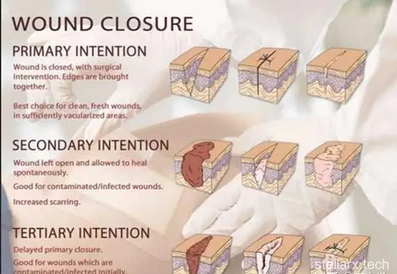
Three ways a wound closes
Primary intention — the classic clean surgical incision or a neat knife cut. Edges are fresh, blood supply good, contamination low, and the two sides are brought together. Only a narrow gap of skin needs to "bridge over," so there is little granulation tissue, a short inflammation phase, and neatly arranged collagen: fast healing, thin scar.
Secondary intention — the wound cannot be closed directly: large tissue loss, infection, contamination, necrosis, missing skin. The body must grow granulation tissue from the bottom up, contract the wound via myofibroblasts, and finally let epithelium crawl in from the edges. Like filling a pothole, then paving it: slow, wide scar, higher infection risk.
Tertiary intention (delayed primary closure) — the surgeon's compromise. The wound starts too dirty, swollen or infected to close safely. It is left open through debridement, drainage, dressing changes and observation, until bacterial load drops and tissue is healthy — then closed late.
The biology of repair
The moment a wound appears, platelets and the clotting system form a fibrin clot. That clot is not waste — it is a temporary scaffold: it stops bleeding and gives leukocytes, keratinocytes, fibroblasts and endothelial cells a route to migrate in.
Then the inflammation phase: neutrophils and macrophages clear bacteria, necrotic tissue and debris. Clear it poorly and infection remains; keep inflammation going too long and the wound stays trapped in "cleaning the battlefield," unable to enter repair.
In the proliferation phase, angiogenesis brings oxygen and nutrients, fibroblasts make collagen, granulation tissue fills the defect, and epidermal cells migrate across the surface. In remodeling — lasting months or longer — loose early collagen is replaced by stronger type I collagen, cross-linked and realigned. Even a well-healed wound usually only reaches about 80% of the original tissue's strength.
Why the finger heals fast and the shin doesn't
A small cut on your finger often looks healed within days because most are shallow lacerations: clean edges, little tissue loss, rich blood supply. As long as there is no deep contamination, tendon damage or foreign body, epithelium crosses the narrow gap quickly.
Shin wounds — especially the pretibial area, lower leg and ankle — are the chronic-healing poster child: thin skin, little subcutaneous tissue, far from the heart, poor venous return that causes edema, poor arterial supply that starves tissue of oxygen. Edema increases the distance between tissues, making oxygen and nutrient diffusion harder. Walking, standing and friction constantly pull at the new tissue.
So two identical-looking 1 cm cuts can be entirely different projects: one is a shallow crack, the other is a small construction site combining poor blood supply, pressure, contamination, tissue loss and tension. Hypoxia, pressure, edema, infection, maceration and dehydration are all documented local factors; diabetes, malnutrition, smoking, obesity, hormonal and immunosuppressive drugs slow healing systemically.
Tension: the factor everyone underestimates
Tension is not how big the wound is — it is whether the two edges are being pulled apart. The chest, back, shoulders, joints and front of the shin are naturally under skin stretch. If a wound is sutured tight, microvessels get compressed, local blood supply drops, oxygen falls, and both collagen synthesis and infection resistance suffer. Tension also keeps stimulating fibroblasts and myofibroblasts, producing more collagen — wider, harder scars, sometimes hypertrophic scars or keloids.
That is why surgeons insist on "tension-free closure": layered suturing, tension-relieving stitches, flaps, grafts, splinting the joint, reducing pull — all so the skin edges meet while blood supply stays intact.
Infection: bacteria count, not presence
Skin is always carrying bacteria. What matters is quantity, virulence, whether necrotic tissue/foreign bodies/dead space give them shelter, and host resistance. WHO's safe surgery framework sums the risk as: bacterial contamination dose × virulence ÷ host resistance. Retained necrotic tissue, suture foreign bodies, drains and remote infections all amplify it.
This is why a dirty wound cannot simply be "sewn shut and done." If dirt, splinters, glass, animal bites, necrotic tissue or pus get sealed inside, the surface may close while an abscess grows beneath. Debridement, irrigation, drainage, removing foreign bodies, controlling blood sugar and antibiotics where needed are not about making the wound "look clean" — they let inflammation end, granulation grow, epithelium cover. Chronic wounds often host biofilms: bacteria wrapped in a protective matrix, far harder to control with routine dressing changes and antibiotics.
Age and body state: the construction crew
Young, well-nourished, well-perfused people — no diabetes, no immunosuppression — have smoother angiogenesis, collagen synthesis, leukocyte killing and epithelial migration. Older skin is thinner, with less dermal collagen and slower epidermal turnover, often combined with atherosclerosis, venous insufficiency, diabetes, anemia, kidney disease or long-term medication.
Protein deficiency weakens fibroblast proliferation, collagen synthesis and angiogenesis. Vitamin C participates in collagen formation. Anemia or low perfusion starves the wound of oxygen. Smoking reduces oxygen delivery through vasoconstriction and carbon monoxide.
When to stop waiting
Rising redness, pain, heat and swelling; pus or odor; wound turning white/yellow/black; expanding size or depth; bleeding you cannot stop; fever and chills; worsening pain; wound dehiscence; sutures or staples coming off early — see a doctor. The American College of Surgeons lists these as wound warning signs.
For diabetic foot wounds, long-standing leg wounds, animal or human bites, deep puncture wounds, contaminated wounds, joint-area wounds, wounds with numbness or limited movement — do not judge by "it scabbed over." A scab is only a dry surface crust; it says nothing about deep repair. Many wounds heal better kept clean, moist, decompressed and protected than left to repeatedly dry and crack.
The hidden causes of a wound that never heals
A wound that stays open past its expected time — or keeps recurring, oozing, bleeding, with a raised/everted/hard edge, abnormal granulation, sinus tracts or odor — deserves a second look behind "weak constitution."
Tuberculosis and atypical mycobacteria: slow-growing organisms cause chronic granulomatous inflammation. Wounds look like ordinary infection but ordinary antibiotics fail. Chronic, atypical, treatment-resistant skin ulcers should include skin TB in the differential; diagnosis needs biopsy for histopathology, acid-fast stain, mycobacterial culture and PCR — not routine bacterial culture.
Malignancy: some "wounds" were never wounds. Tumor invasion creates malignant fungating wounds that never follow the normal healing sequence. And long-standing scars can turn malignant — Marjolin ulcer, most often squamous cell carcinoma. Old burn scars, pressure ulcers, chronic leg ulcers and osteomyelitis sinuses that change edge, bleed, smell or grow abnormal granulation warrant a low biopsy threshold; guidelines suggest biopsy for leg wounds with no improvement after 4–12 weeks of proper care.
Radiation injury: the "3H" tissue — hypoxia, hypovascularity, hypocellularity. Irradiated skin is ischemic, fibrotic and cell-poor, so repair is permanently impaired.
Liver failure / cirrhosis with low albumin: the liver makes albumin, clotting factors and complement. Ascites presses on abdominal wounds from inside — constant high tension, moisture, leakage, contamination risk.
Alcoholism: acute alcohol disrupts immune cell function; chronic use brings protein/vitamin deficiency and liver damage. Alcohol weakens all three construction crews — immune cells, endothelial cells, fibroblasts.
Anemia and hypoxia: oxygen drives collagen hydroxylation, fibroblast activity, angiogenesis, epithelial migration and leukocyte killing. But mild anemia alone does not doom healing — tissue oxygenation depends on hemoglobin, lung function, cardiac output, local flow, microcirculation and edema together.
The next time a "small cut" refuses to heal, the question is not how big it is. It is which stage of the repair project stalled, and why the local or systemic conditions let it stall.
Curious what the scaffold that builds every healed scar looks like? Fibrous connective tissue specimens show the dense, parallel collagen fiber bundles that give a repaired wound its strength — under the microscope you can see exactly why remodeling matters. Search for WWAI in your app store: its Fibrous connective tissue specimen lets you zoom through the collagen architecture on screen.

Related reading

Tetanus From a Tiny Wound: No Rust Needed

Vibrio vulnificus: Seafood Prick Danger
References
Brunicardi F.C. et al. Schwartz's Principles of Surgery, 11th ed., Chapter 9 "Wound Healing." McGraw-Hill, 2019.
Guo, S. & Dipietro, L.A. "Factors affecting wound healing." Journal of Dental Research 89(3), 2010: 219-229.
Ozgok Kangal, M.K. & Kopitnik, N.L. "Physiology, Wound Healing." StatPearls, 2025.
World Health Organization. WHO Guidelines for Safe Surgery 2009: Safe Surgery Saves Lives. Geneva: WHO.
American College of Surgeons. Surgical Wound Care.
- Resistant Starch and Fatty Liver: What 40g a Day Really Does
- 抗性澱粉能救脂肪肝嗎?每天40克的數據與真相
- 大水理論、薄肌理論其實是好建議?最便宜的養生法最有科學依據
- The 'Big Water, Thin Muscle' Meme Is Quietly Good Advice
- Bananas Come in Every Color of the Rainbow
- The Bacterium That Lived Alone for a Million Years
- What ICU Actually Feels Like: Held Between Life and Death
- Why Some Wounds Heal in Days and Others Never Do
- The Heaviest Insect Ever Measured Is Back From the Brink
- Your Gut's "Second Brain": 500 Million Neurons of Its Own
全部评论