Ectopic Tooth in Eyelid: A World-First Superior Orbit Case
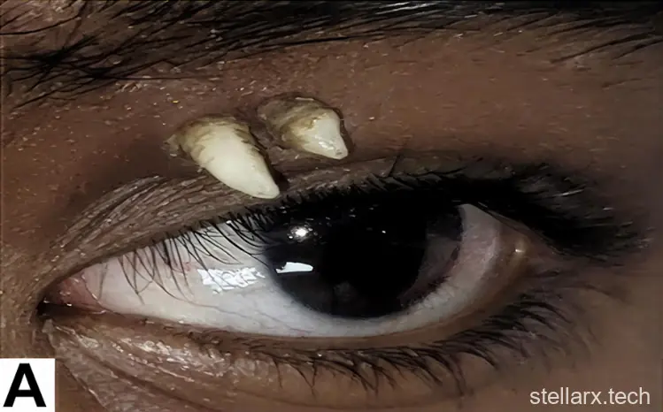
A 16-year-old boy in India walked into a hospital with a month-old, painless swelling in his left upper eyelid. No trauma. No discomfort. Just a quiet bump that had been sitting there — until doctors pulled back the lid and found two hard, white points that looked exactly like teeth.
They were. One canine tooth, fully formed, had grown inside the top of his eye socket and pushed two of its cusps through the thin skin below the brow. The case, reported by Meghana Tanwar and colleagues at Aravind Eye Hospital in Madurai, was published in Ophthalmology — and according to the authors, it is the first and only ectopic tooth ever documented in the superior orbit.
Two White Points That Felt Like Teeth
On examination, the two projections sat at the inner side of the eyelid. They had a fairly smooth surface, were hard to the touch, and were well defined at the edges. "Suggestive of a tooth," the team wrote.
Because the swelling had appeared with no injury, the initial guesses were a foreign body or a teratoma — a rare tumor that can grow fully formed tissues, including teeth, hair, or bone. In this boy's case, a clue came early: the two white spikes were not two separate teeth. They were the two visible cusps of a single tooth, poking through skin that is only a few millimeters thick.
A CT Scan Settled It
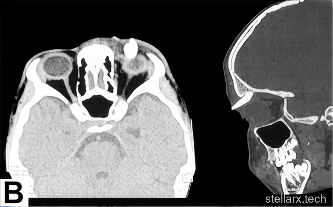
Computed tomography showed a dense structure with a complete crown-and-root morphology arising from the left orbital roof and extending toward the eyelid. "The structure of a lone tooth in the superior orbit was quite obvious," Tanwar told the media.
The location is what makes the case extraordinary. The roof of the orbit is a thin plate of bone that separates the eye socket from the front of the brain — far from the tooth-forming tissues of the upper jaw. In the mouth, permanent upper canines normally erupt around ages 11 or 12. How one came to form in a bone that has no business making teeth is a question the report does not answer.
A Mature Canine, Far From Home
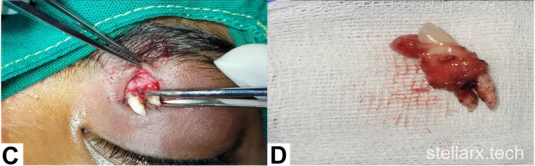
The tooth was removed surgically, with meticulous dissection of the tooth and its immediate surrounding tissue. The procedure took about 20 minutes under general anesthesia, after consultation with a maxillofacial surgeon. At the two-week postoperative visit the wound had healed well, and at six months the patient had a minimal scar and no problems.
Laboratory examination identified the specimen as a mature canine tooth with no features of a teratoma. The boy's problem was mainly cosmetic rather than functional — the tooth was not pressing on the eye, muscles, or nerves — though the authors noted that if it had migrated toward those structures, mechanical compression could have caused functional issues.
What Is an Ectopic Tooth?
An ectopic tooth is a tooth that develops and erupts outside the normal dental arch. The mechanism is not fully understood; leading hypotheses involve abnormal embryonic development, displacement of the tooth germ, trauma, and infection. A 2023 review of risk factors added developmental disorders, pathological conditions, genetic factors — and even "developmental atavism," an evolutionary throwback.
How rare is that? A 2023 review in Imaging Science in Dentistry searched the literature from 1980 to 2022 and found 61 reported ectopic teeth. About 61% were in the maxillary sinus, the air space inside the cheekbone — the most common hideout by far. Ectopic first permanent molars, meanwhile, affect roughly 2% to 6% of people, but those stay inside the mouth.
Teeth Have Turned Up in Strange Places Before
Teeth have been found in the nose — one 38-year-old man had an ectopic tooth inside his nostril after years of breathing problems — in the chin, the palate, and, in a 2011 image in the New England Journal of Medicine, inside the maxillary sinus. A 2019 report described a 30-year-old man with a cyst growing from an impacted tooth in the orbital floor.
The eyelid itself is a different story. Since the first report in 1934, only a handful of eyelid ectopic teeth have been described — just four cases including this one — and all were in the lower eyelid. In 2019, Liu and colleagues reported a Chinese infant with a congenital mass in the left lower eyelid that contained a tooth. Tanwar's own team had previously removed an odontogenic choristoma — normal dental tissue in an abnormal location — from the floor of the orbit of a seven-year-old girl.
When a Tooth-Shaped Mass Is Not an Ectopic Tooth
A tooth-shaped finding on imaging is not automatically an ectopic tooth. The differential is a genuine clinical puzzle:
Orbital teratoma. In 2013, Chawla and colleagues reported a seven-year-old girl with a congenital right orbital mass and an absent eye. Pathology showed tissues from all three germ layers — a mature orbital teratoma.
Odontogenic choristoma. In 2023, Theiveegan and Tanwar's colleagues reported a seven-year-old girl whose MRI suggested a mature teratoma in the right orbit, but whose post-surgical pathology revealed an odontogenic choristoma containing a complete molar.
Dentigerous cyst. A 2025 review in Case Reports in Ophthalmology tabulated seven orbital tooth cases from 1887 to 2023, including a 21-year-old woman with an ectopic canine hidden inside a cyst in the inner orbit — a condition that mimicked a lacrimal sac infection.
The two 2023 Orbit cases both came from Aravind Eye Hospital — the same institution that treated the 16-year-old. Tanwar was second author on the 2023 case and first author on this one. The thread runs through one hospital's experience with teeth that refuse to stay put.
The Bigger Picture
For patients, the practical lesson is simple: any persistent swelling or lump around the eye, even a painless one, deserves a check. Imaging settled this case in minutes; surgery removed it in twenty. For clinicians, the takeaway is that an unusual, hard mass near the eye can occasionally have a dental origin — and that a tooth-shaped structure is not always what it looks like under the microscope of differential diagnosis.
For everyone else, it is a reminder that the body's tissues are not always where they belong — and that the enamel on your teeth began as oral epithelium, the same kind of cell you can scrape off the inside of your cheek.
Want to see what tooth-forming tissue looks like up close? The cells that give rise to tooth enamel start out as ordinary epithelial cells — the same flat, nucleus-bearing cells you can observe in an Oral Epithelium specimen under a virtual microscope, where the purple cell nuclei are clearly visible. Just search "WWAI" in your app store to bring a digital microscope to your pocket and look at the microscopic world anytime you're curious.
Related reading

Dry Eyes? The Problem Is Oil, Not Water

Eye Stroke: 90-Minute Window That Saves Sight

Plant Bristles in Your Eye: Why Not to Rub
References
Tanwar M, Sharma V, Kim U. Ectopic canine tooth in the superior orbit. Ophthalmology. 2026;133(6):e84. DOI: 10.1016/j.ophtha.2025.12.020.
Arora P, Nair MK, Liang H, et al. Ectopic teeth with disparate migration. Imaging Science in Dentistry. 2023;53(3):229-238. DOI: 10.5624/isd.20230040.
Liu J, Chen J, Zhang YS, et al. A tooth lesion in the eyelid in a Chinese infant. Int J Ophthalmol. 2019;12(11):1807-1808. DOI: 10.18240/ijo.2019.11.22.
Chawla B, Chauhan K, Kashyap S. Mature orbital teratoma with an ectopic tooth and primary anophthalmos. Orbit. 2013;32(1):67-69. DOI: 10.3109/01676830.2012.747212.
Theiveegan D, Tanwar M, Kulkarni P, et al. Orbital tooth — not always a teratoma. Orbit. 2023;42(6):650-653. DOI: 10.1080/01676830.2022.2072900.
Two white spikes broke through the skin above a teen's eye, and surgeons removed a canine tooth. Medical Daily. 2026-09-29. https://www.medicaldaily.com/canine-tooth-eye-socket-teen-eyelid-479244
Doctors remove tooth growing between teen's eyelid and eyebrow. Juta MedicalBrief (citing MedPage Today). 2026-01-28. https://www.medicalbrief.co.za/doctors-remove-tooth-growing-between-teens-eyelid-and-eyebrow/
Ectopic tooth found in upper eye socket thought to be world first. Dentistry.co.uk. 2026-10-14. https://dentistry.co.uk/2026/10/14/ectopic-tooth-found-in-upper-eye-socket-thought-to-be-world-first/
Dentigerous cyst masquerading as lacrimal sac diverticulitis. Case Reports in Ophthalmology. 2025;16(1):95-105. https://karger.com/cop/article/16/1/95/919025
- Sweet Potato Leaves: Fiber, Calcium, Lutein in One Green
- 地瓜葉的營養價值:鈣比牛奶高、葉黃素超西蘭花
- 蟑螂粉的真相:71% 粗蛋白不是蛋白
- Why Cockroach "Protein Powder" Isn't Protein
- Autocatalysis in Evolution: From Fish to Humans
- 自催化的演化:從魚到人的 5 億年
- Ectopic Tooth in Eyelid: A World-First Superior Orbit Case
- 眼瞼長牙的真相:全球首例眶上異位牙
- What Is Lean Muscle? 4 Factors Behind 'Thin Muscle' Physique
- 薄肌理論是啥?4 個特徵決定你的肌肉線條

全部评论